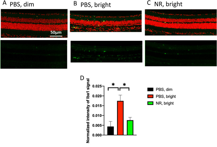
Figure 8.
NR treatment prevents activation of Iba1-positive cells after induction of LIRD. Both PBS- and NR-treated BALB/c mice received toxic light exposure and were euthanized at 1 week after exposure. (A–C) Representative morphologic images of each group; green is Iba1 immunofluorescence signal and red is PI staining. (D) Iba1-positive signals were quantified from the entire retina. NR treated mice (green bar) exhibited significantly less Iba1 signals compared with the PBS treated group (red bar). *P < 0.05 by one-way ANOVA with Newman-Keuls multiple comparisons post hoc test. N = 4–5 mice per group. Error bars represent SEM. Size marker = 50 µm.

