Fig. 7.
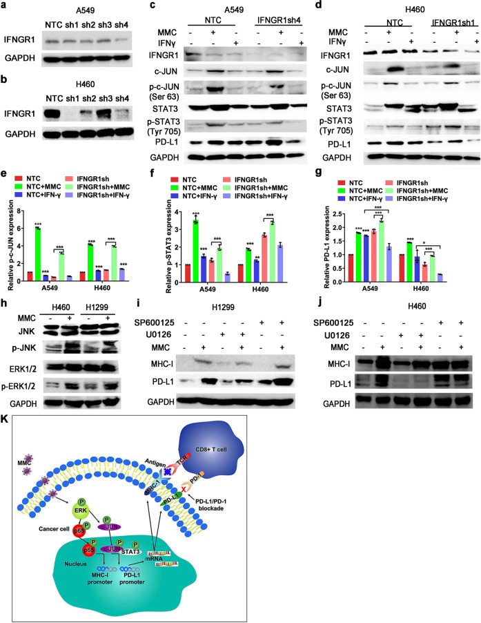
MMC induced MHC-I and PD-L1 expression via ERK signaling. a, b Western blot analysis showing the knockdown of IFNGR1 by four different shRNAs (sh1-sh4) in A549 and H460 cells. c–g Western blot analysis showing the PD-L1 protein expression in IFNGR1-knockdown cells with or without MMC (0.4 μM, 48 h) or IFN-γ (1 ng/mL) treatment. The IFN-γ (1 ng/ml) was used a positive control. The expression level of the protein was normalized with GAPDH. Data are presented as relative ratio to the untreated cells (e–g). *p < 0.05; **p < 0.01; ***p < 0.001. h Western blot analysis showing ERK and JNK protein expressions in MMC treated cells (0.4 μM, 48 h). i, j The PD-L1 and MHC-I expression in MMC treated cells (0.4 μM, 48 h) in the presence of an ERK inhibitor (U0126 (20 μM)) or a JNK inhibitor (SP600125 (15 μM)). The cells were pretreated with U0126 or SP600125 for 3 h. The expression level of the protein was normalized with GAPDH. *p < 0.05, **p < 0.01, ***p < 0.001. k A schematic diagram summarizing the molecular mechanisms underlying the upregulation pf PD-L1 and MHC-I by MMC in NCSLC cells. MMC activates the ERK pathway, which subsequently enhances the binding of c-JUN to the PD-L1 promoter and recruits its co-factor STAT3 to increase PD-L1 expression. The upregulated ERK pathway also activates p65 to increase the MHC-I expression. The up-regulations of PD-L1 and MHC-I then induce more and more CD3+ and CD8+ T-cell infiltration in tumor tissue and kill the tumor cells

