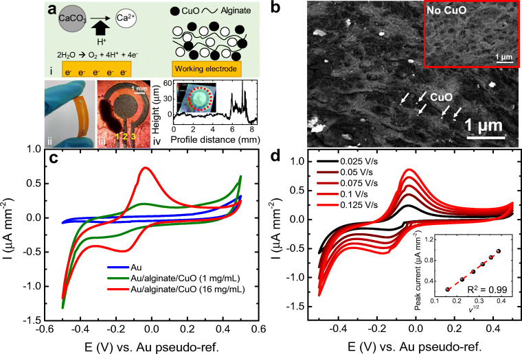
Fig. 1.
(a) Alginate electrodeposition with CuO nanopowder. (i) Scheme of electrodeposition mechanism. The flexible chip is shown in (ii), with the membrane formed after 140 s electrodeposition time. Yellow numbers indicate electrode function: (1) working, (2) counter, (3) pseudo-reference. Film thickness (10 μm) is shown in (iv), measured along the red dotted circle in inset. (b) SEM imaging of alginate membrane with CuO NPs. Backscattered electrons provide the contrast, with the white spots belonging to the CuO (arrows indicate some examples). Inset shows the membrane without CuO, where no white spots can be seen. (c) Cyclic voltammogram of bare electrodes (blue line) and modified electrodes (red and green), showing the appearance of the redox pair belonging to CuO. (d) Scan rate test and calibration (inset) showing linear relation with square root of the scan rate. All cyclic voltammetries were performed in imidazole buffer at pH 7.4 and room temperature

