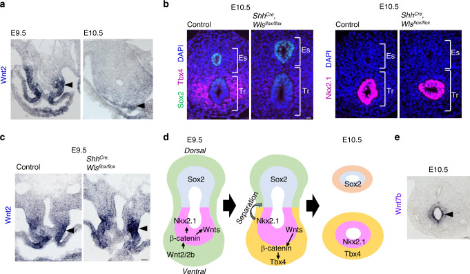
Fig. 3. Endodermal Wnt ligands induce Tbx4 expression for tracheal mesoderm development of mouse trachea.
a In situ hybridization for Wnt2 mRNA during tracheoesophageal segregation. Arrowheads indicate Wnt2 expression in the ventrolateral mesoderm at E9.5 and E10.5. n = 2/2 embryos. b Transverse sections of ShhCre, Wlsflox/flox mouse embryos and littermate controls at E10.5. Left panels show sections stained with Sox2 (green), Tbx4 (magenta), and DAPI (blue). Right panels show sections stained for Nkx2.1 (magenta) and DAPI (blue). n = 3/3 embryos per genotype. c In situ hybridization for Wnt2 mRNA in ShhCre, Wlsflox/flox mouse embryos and littermate controls at E9.5. Arrowheads indicate Wnt2 expression in the ventrolateral mesoderm. n = 2/2 embryos. d Refined model of tracheoesophageal segregation and tracheal mesodermal differentiation. e In situ hybridization for Wnt7b mRNA in mouse embryo at E10.5. Arrowhead indicates Wnt7b+ cells. n = 2/2 embryos. Eso Esophagus, Lu Lung, Tr Trachea. Scale bar; 40 μm (a–c), 50 μm (e).

