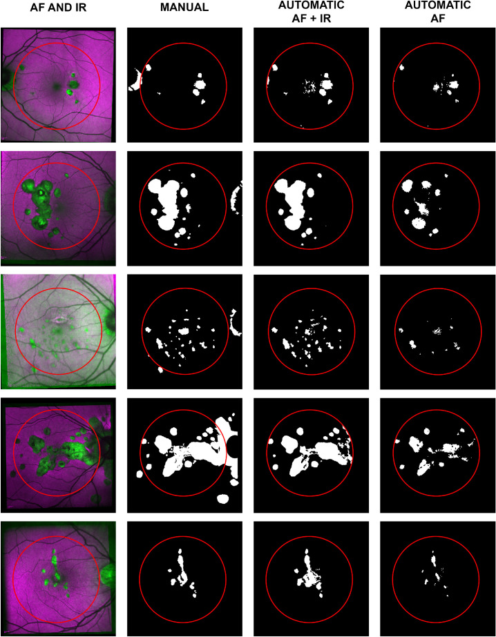
Figure 2.
A random subset of five of the 18 selected eyes. The first column shows a combination of the IR and AF. The second column shows the manual segmentation as a binary map of “0” (non-lesion, in black) and “1” (lesion, in white). The third column shows the automatic segmentation based on IR and AF for the central 22.5°, delimited by a red circle. The fourth column shows the results of the same classification model trained on AF only.

