Image 4.
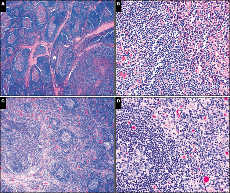
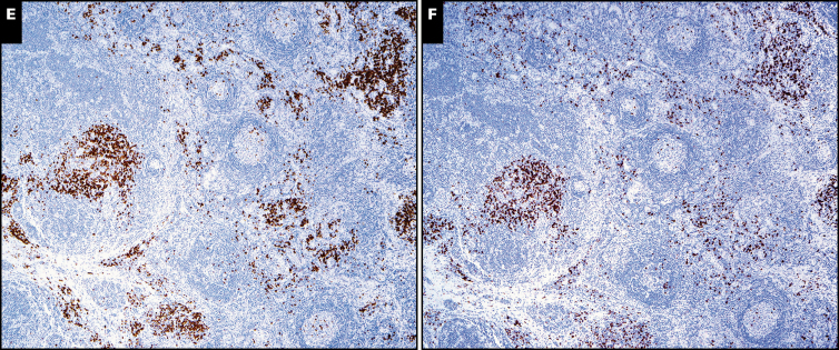
Castleman morphologic features in multicentric cases. A and B, The symptomatic patient with idiopathic multicentric Castleman disease demonstrated the plasma cell (PC) variant by morphology (×20) with reactive germinal centers (A) and numerous interfollicular plasma cells (B, right side of photomicrograph; ×200). C and D, The patient with TAFRO (thrombocytopenia, anasarca, fever, reticulin fibrosis of the bone marrow, and organomegaly) had lymph node morphology demonstrating mildly atrophic germinal centers (C; ×40) with moderate mantle zone “onion skinning” and abundant interfollicular plasma cells (D, right side of photomicrograph; ×200). The κ (E) and λ (F) stains show polytypic plasma cells in the lymph node biopsy from the patient with TAFRO (both at ×40).


