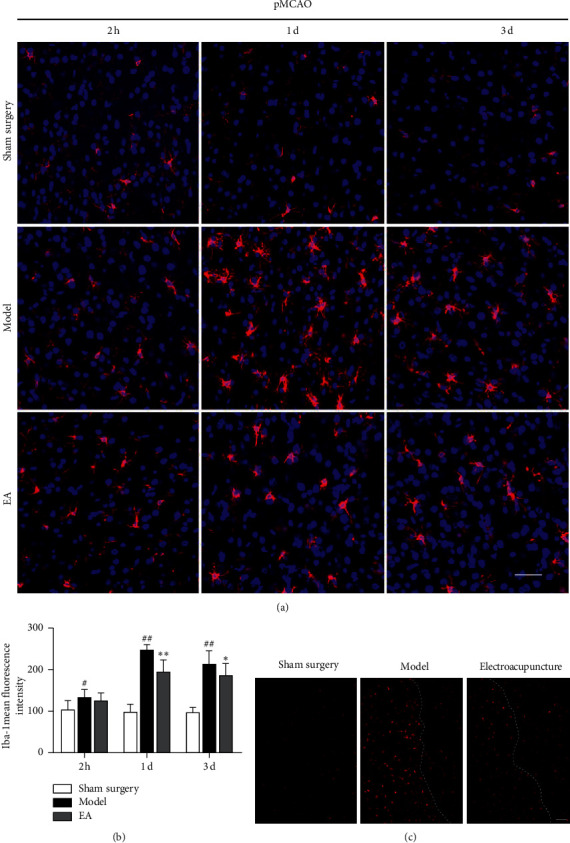
Figure 3.

Expression of Iba-1 in pMCAO rats and the effects of electroacupuncture on the activation of microglia. (a) Expression of Iba-1 in the cortical ischemic penumbra (IP) 2 h 1 d and 3 d after pMCAO, as determined by laser confocal scanning microscopy. Bar = 50 μm. (b) Mean fluorescence intensity (MFI) of Iba-1. The expression of Iba-1 in the model group was increased after cerebral ischemia (#P < 0.05, ##P < 0.01 versus the sham surgery group). One day and 3 d after cerebral ischemia, electroacupuncture decreased the expression of Iba-1 in pMCAO rats (∗P < 0.05 and ∗∗P < 0.01 versus the model group). (c) The expression of Iba-1 in the cortical IP 1 d after pMCAO. Fluorescently labeled microglia were red, and there were areas of the IP in which microglia were obviously activated in the model group. In contrast to that in the model group, the expression of Iba-1 in the electroacupuncture group was reduced. Bar = 100 μm.
