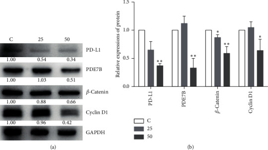
Figure 3.

Analysis expressions of PDE7B, PD-L1, β-catenin and cyclin D1. Following the treatment of 4T1 cells with 25 or 50 μg/ml of EA11, PDE7B, PD-L1, β-catenin and cyclin D1, proteins were assessed by western blot. Values are presented as mean ± SD from three experiments. ∗P < 0.05, ∗∗P < 0.01 vs. C group.
