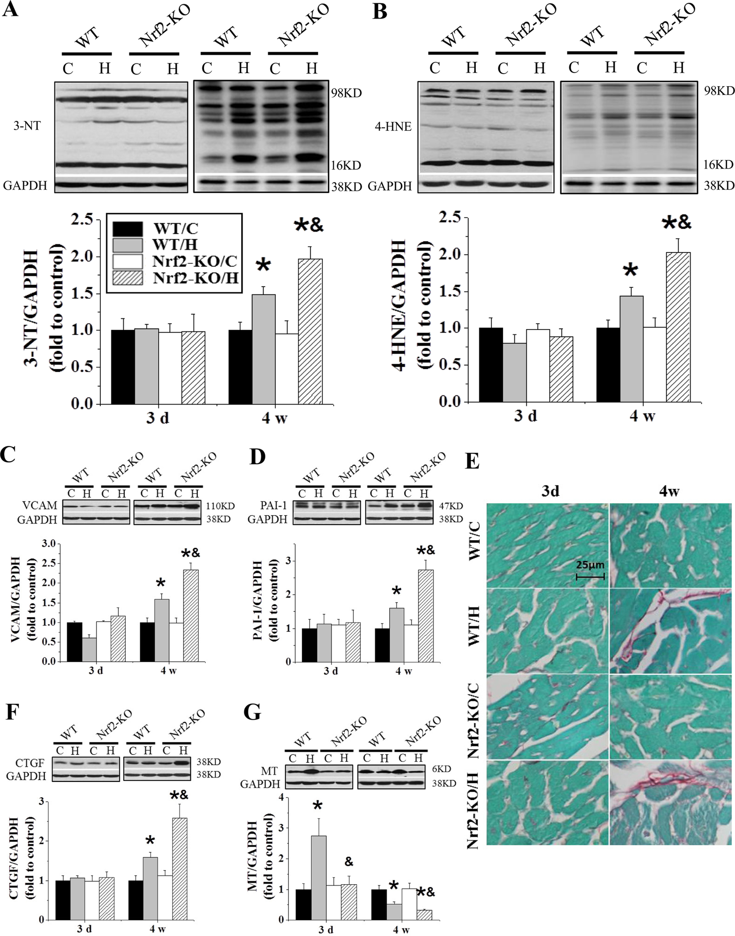
Figure 3. Nrf2-KO mice exacerbate IH-induced oxidative stress, inflammation and fibrosis.

Nrf2-KO and C57BL/6J WT mice were exposed to IH for indicated times. Cardiac oxidative damage was measured by Western blots for 3-nitrotyrosine (3-NT, A) and 4-Hydroxynonenal (4-HNE, B). Cardiac inflammation and fibrosis were measured by Western blots for vascular cell adhesion molecule 1 (VCAM-1, C), plasminogen activator inhibitor-1 (PAI-1, D), and connective tissue growth factor (CTGF, F). Cardiac collagen was measured by Sirius-red staining (E). MT (G) expression was measured by Western blots. Data are presented as mean ± SD (n=5). *, p<0.05 vs WT/C; &, p<0.05 vs WT/H.
