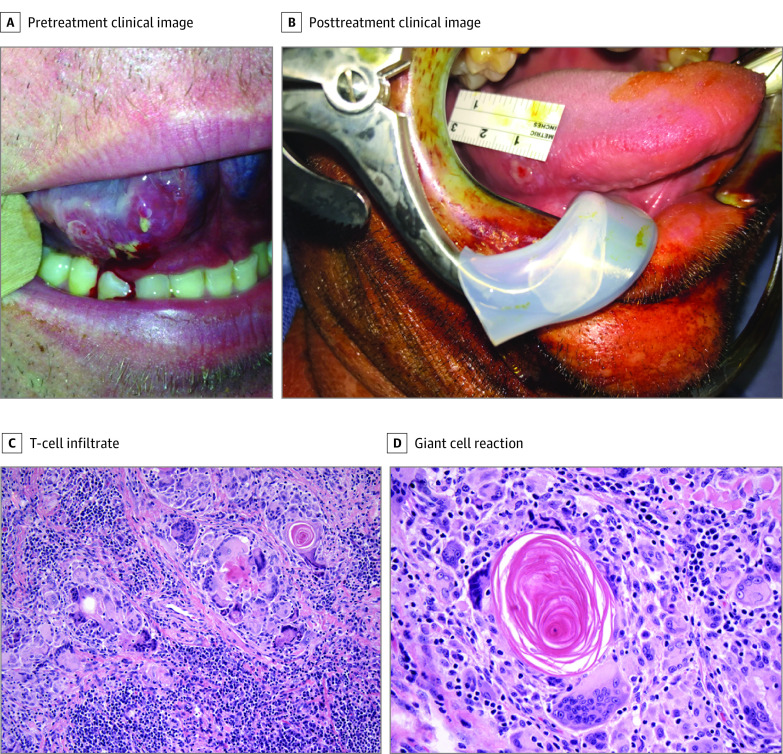
Figure 3. Clinical and Pathologic Features of Response.
A, Pretreatment image shows a cT2 squamous cell carcinoma of the right oral tongue. B, Fifteen days following cycle 1 of preoperative immunotherapy, image at the time of surgery demonstrates near-complete clinical regression of primary lesion (no residual squamous cell carcinoma identified on pathologic analysis). C, Original magnification ×200, and D, original magnification ×400 histopathologic images at the time of surgery following neoadjuvant immunotherapy in another patient with a robust response demonstrate evidence of robust T-cell infiltrate with evidence of regressed tumor, giant cell reaction, and acellular keratin. C and D, Hematoxylin-eosin.

