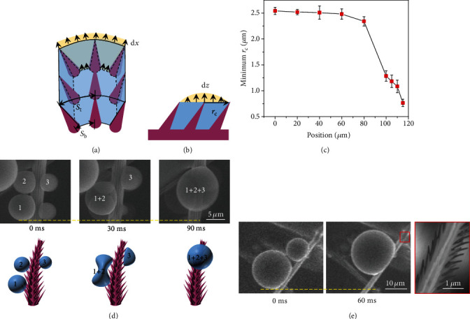
Figure 2.

Microscopic condensation dynamics. (a) Schematic image showing the extension of liquid in the lateral direction by an incremental distance dx. Sb and St represent the space between two neighboring setae at the bottom and apex of nanoratchets. (b) Schematic image showing the extension of liquid along vertical direction by an incremental distance dz. rc indicates the base area of condensate droplet. As the vapor-to-liquid phase change proceeds continuously, condensate embryo nucleating within the nanoratchet arrays extends laterally, until it grows large enough to be confined by the underlying nanostructure. (c) The minimum base area of droplet as a function of position. Here, position 0 μm represents the bottom of setae. The error bars are the standard deviation of the measurements. (d) ESEM images and their corresponding schematic images representing the continuous propagation of multiple droplets in a step-by-step manner. (e) ESEM images showing the directional motion of droplet enabled by the coalescence with adjacent droplet. Remarkably, condensate droplets always move towards the tip of seta even in the case when small droplet merges with large droplet. The picture with red border demonstrates the direction of the underlying substrate.
