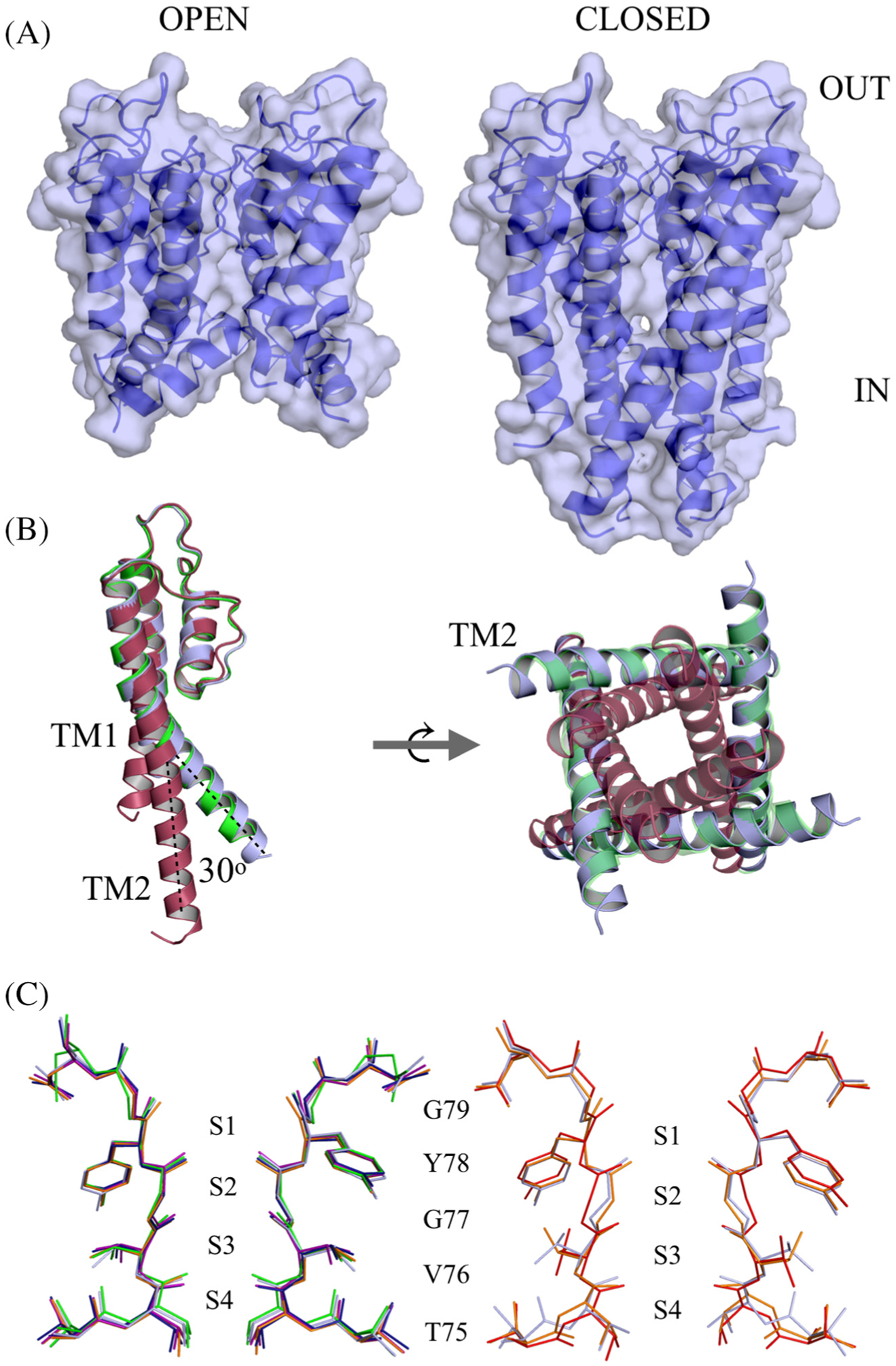
Figure 1.

Crystal structure of open and closed-gate wt KscA soaked in BaCl2. (A) Sideview, space-filling model of the open-gate conformation (left) and the closed-gate conformation (right) of wt KcsA. The channel is oriented with the extracellular side on top. (B) Superposition of open-gate wt KcsA (blue) on 3F7V open mutant (green) and 1K4C closed-conductive channel (red). One monomer is shown for clarity; the dotted lines illustrate the rigid body movement of TM2 hinged at Ser102 (left). A cytoplasmic view of the tetramer showing the inner gate of the channel (right). (C) Comparison of selectivity filter of wt KcsA open-gate structure soaked in 1, 2, 4, 5 or 10 mM Ba2+ (left). Superposition of the selectivity filter open-gate wt KcsA (blue) with the selectivity filter of the conductive 1K4C (red) and the constricted 1K4D (orange) structures (right).
