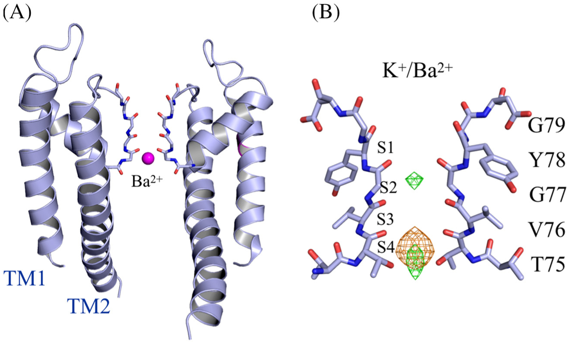
Figure 2.

Ba2+ binding in the open-gate wt KcsA structures. (A) Side view of the open-gate wt KcsA soaked in 1 mM BaCl2/5 mM KCl. Two diagonally opposing monomers are shown for clarity. Ba2+ (magenta sphere) is shown binding in the selectivity filter. (B) Close view of the selectivity filter in the region Thr74–Asp80. Anomalous Fourier difference map (orange mesh) for Ba+2 contoured at 8 σ, and Fo − Fc map (green mesh) contoured at 6 σ.
