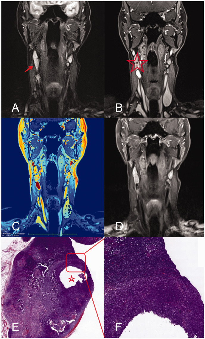
Fig. 2.
Representative images of a patient with non-keratinizing squamous cell carcinoma and a metastatic cervical LN with a short-axis diameter of 11 mm. Metastatic LN (arrow) on coronal images from T2W TIRM (a), 3D DCE (b); 3D DCE derived pixel-by-pixel color-coded map of the iAUC (red = maximum, blue = minimum) (c); contrast-enhanced T1W imaging (d); histological analysis (e, H&E staining; overview); close-up of metastatic LN (f). The hypoperfused area (asterisk) is delineable in the 3D DCE sequence only (asterisk) and confirmed histologically. DCE, dynamic contrast-enhanced; iAUC, initial area under the curve; LN, lymph node; T1W/T2W, T1-/T2-weighted.

