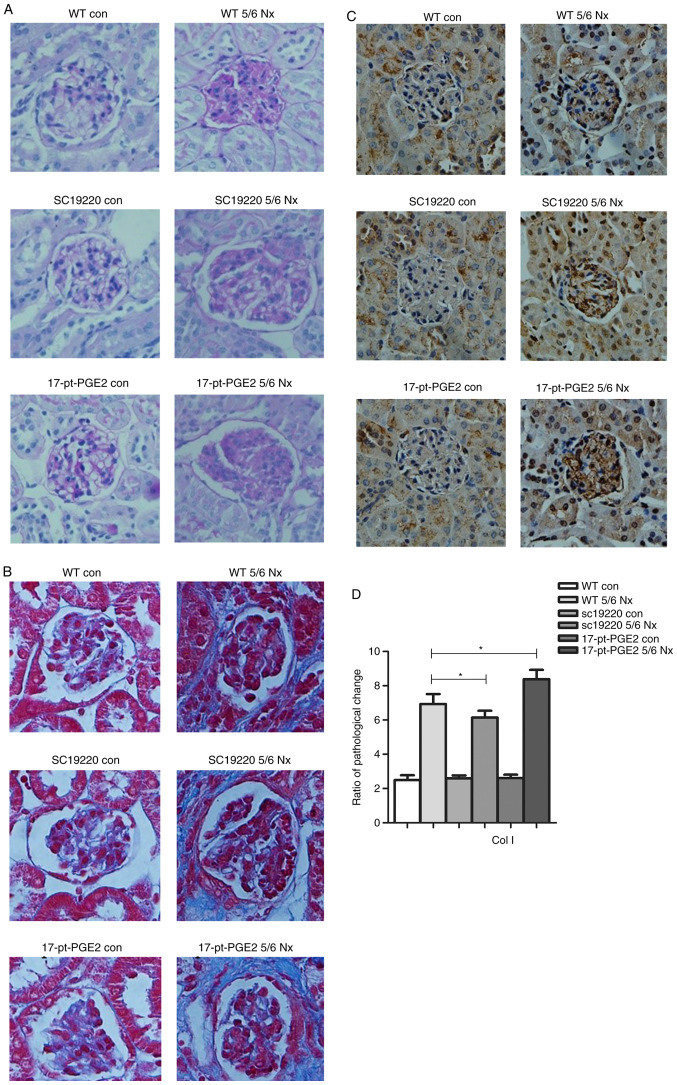
Figure 2.
Effects of blocking or stimulating the prostaglandin E2 receptor 1 on the degree of renal fibrosis. Images were captured with a Hitachi VKC150 camera. (A) Periodic acid-Schiff staining was used to visualize the accumulation of extracellular matrix. (B) Masson's staining was used to visualize collagen deposition. WT, wild-type; 5/6 Nx, five-sixths nephrectomy; con, control; 17-pt-PGE2, 17-phenyl-trinor-prostaglandin E2 ethyl amide. Effects of blocking or stimulating the prostaglandin E2 receptor 1 on the degree of renal fibrosis. Images were captured with a Hitachi VKC150 camera. (C) Immunohistochemistry was used to visualize the expression of Col1 (magnification, ×400). (D) Semi-quantitative analysis of Col1 expression. *P<0.05. WT, wild-type; 5/6 Nx, five-sixths nephrectomy; Col1, Human collagen type 1; con, control; 17-pt-PGE2, 17-phenyl-trinor-prostaglandin E2 ethyl amide.

