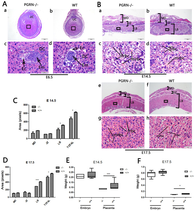
Figure 2.
Changes in PGRN−/− mouse placentas and embryos. (A) H&E staining of (a and c) PGRN−/− sections and (b and d) WT (C57) embryos on E6.5. (c and d) Higher magnifications of the boxed areas in (a) and (b) show the embryos. (B) H&E staining of PGRN−/− sections and WT (C57) placentas on (a-d) E14.5 and (e-h) E17.5. Higher magnifications of the boxed areas in (a), (b), (e) and (f) show the structure of the labyrinth and the red blood cells of PGRN−/− and WT placentas on the maternal and fetal sides on (c and d) E14.5 and (g and h) E17.5. (C) Morphometrical analysis of sections in (Ba and b) E14.5. (D) Morphometrical analysis of sections in (Be and f) E17.5. (E) Weights of placentas and embryos from WT and PGRN−/− mice on E14.5 were analyzed. (F) Weights of placentas and embryos from WT and PGRN−/− mice on E17.5 were analyzed. Scale bar, 500 µm; magnified scale bar, 50 µm. *P<0.05, ***P<0.001. PGRN, progranulin; H&E, hematoxylin and eosin; -/-, PGRN−/− mice; +/+, WT mice; WT, wild-type; E, embryonic day; Em, embryo; MD, maternal decidua; JZ, junctional zone; LB, labyrinthine layer; frbc, fetal red blood cells; mrbc, maternal red blood cells.

