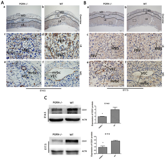
Figure 3.
CD31 expression in developing placentas from PGRN−/− and WT mice on E14.5 and E17.5. Immunohistochemical detection of CD31 in the serial placental sections on (A) E14.5 and (B) E17.5. (A and B) Boxed areas in (a) are magnified in panels (c) and (e). Boxed areas in (b) are magnified in panels (d) and (f). (C) Western blot analysis and quantification of the results in placentas on E14.5 and E17.5. Scale bar, 500 µm; magnified scale bar, 50 µm. *P<0.05. CD31, cluster of differentiation 31; PGRN, progranulin; -/-, PGRN−/− mice; WT, wild-type; E, embryonic day; MD, maternal decidua; JZ, junctional zone; LB, labyrinthine layer; MBS, maternal blood sinus; FBV, fetal blood vessel; VEC, vascular endothelial cells; mrbc, maternal red blood cells; ACTB, β-actin.

