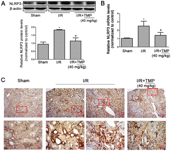
Figure 3.
TMP reduces the expression of NLRP3 in renal tissues from renal I/R rats. (A) Representative western blotting image and summarized data showing the expression of NLRP3 in the kidney tissues of rats in the Sham, I/R and I/R+TMP groups. (B) Reverse transcription-quantitative PCR was used to detect NLRP3 mRNA expression in the kidney tissues of rats in each group. (C) Immunohistochemical staining showing the expression of NLRP3 in renal tissues in each group. The red arrows indicate the positive expression of NLRP3 in infiltrating inflammatory cells. Red boxes highlight the corresponding area of the high-power images. *P<0.05 vs. Sham rats; #P<0.05 vs. I/R rats. I/R, ischemia-reperfusion; TMP, tetramethylpyrazine; NLRP3, NACHT, LRR and PYD domains-containing protein.

