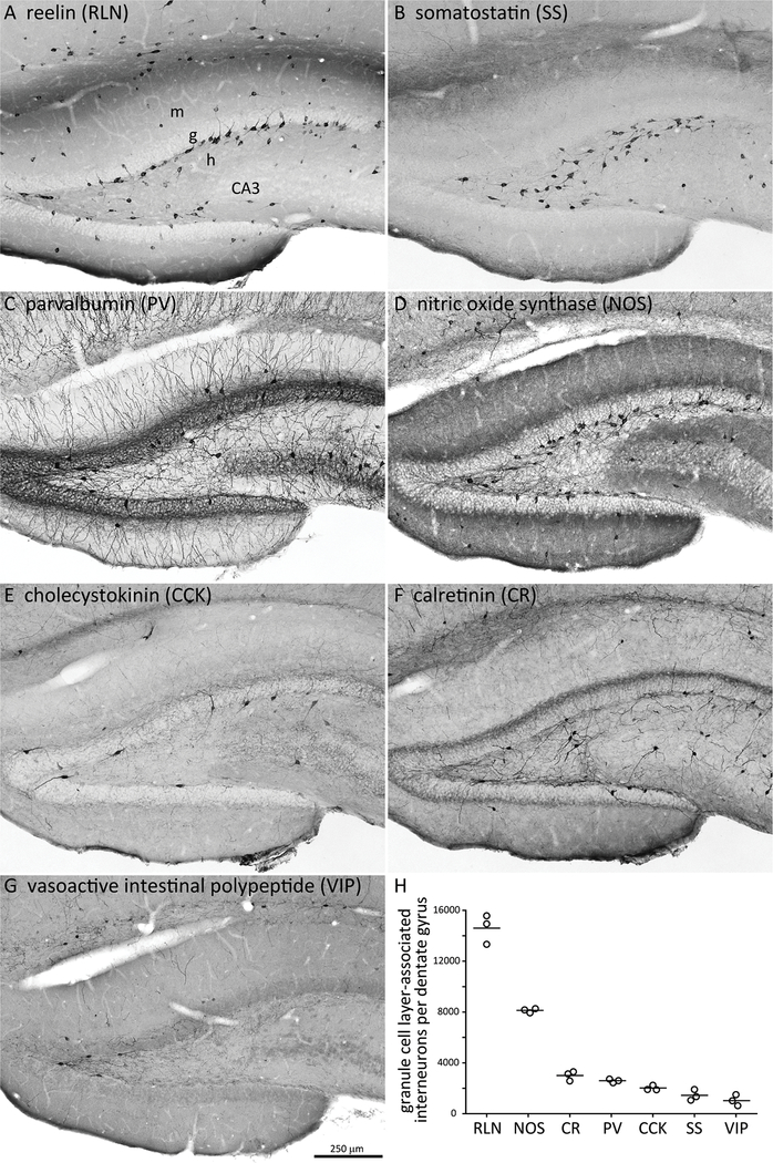
Figure 4.
Cells that express reelin or nitric oxide synthase account for most granule cell layer-associated interneurons. A-G Adjacent hippocampal sections from a control rat processed for different interneuron markers. m = molecular layer, g = granule cell layer, h = hilus. H Number of interneurons in or adjacent to the granule cell layer per hippocampus. Circles represent individuals, lines indicate averages.

