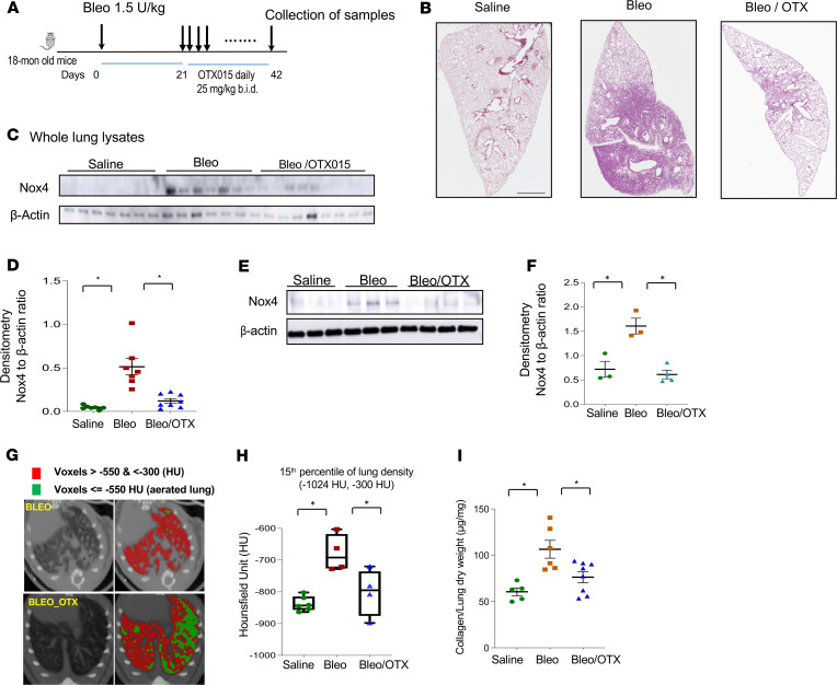
Figure 7. The inhibitor OTX015 facilitates resolution of established lung fibrosis in aged mice.
(A) The schedule of lung fibrosis induced by bleomycin injury in 18-month-old mice. OTX015 was given twice daily orally from day 21–42 after bleomycin injury. All the mice were sacrificed at day 42 after bleomycin injury. (B) Whole lung histology by immunohistochemistry with H&E staining of mice with saline, bleomycin injury (bleo), or bleomycin injury with OTX015 treatment (Bleo/OTX). (C) Mouse whole lung tissue lysates from saline (n = 7), bleomycin (n = 7), and bleomycin with OTX015 treatment (n = 9) groups were subjected to SDS-PAGE and Western blots analysis for Nox4 and β-actin. (D) Densitometry of the Western blots in C for the ratio of Nox4 to β-actin (mean ± SD). (E) Primary lung fibroblasts were cultured from mouse lungs. Whole cell lysate from saline (n = 3), bleomycin (n = 3), and bleomycin with OTX015 treatment (n = 4) groups were collected at passage 1 and subjected to SDS-PAGE and Western blots analysis for Nox4 and β-actin. (F) Densitometry of the Western blots in E for the ratio of Nox4 to β-actin in the primary murine lung fibroblasts (mean ± SD). (G) Representative axial micro-CT images of mouse lungs 6 weeks after they were subjected to bleomycin injury with/without OTX015 treatment. Voxels of the lung field that are below –550 Hounsfield units are in green (representing aerated lung) and those between –550 and –350 Hounsfield units are in red (representing nonaerated lung). (H) Quantitation of lung density in uninjured (saline) and injured (Bleo) mice treated with/without OTX015. Whisker plots represent mean ± SD; n = 4–6 per group (saline, n = 6; bleo, n = 4; bleo/OTX015, n = 4). *P < 0.05, by 2-tailed t test. (I) Hydroxyproline content in lungs of mice after saline (n = 5), bleomycin (n = 6), or bleomycin with OTX015 treatment (n = 8) (mean ± SD). *P < 0.05, for comparisons of indicted groups as compared with the bleomycin group, by 2-tailed t test.

