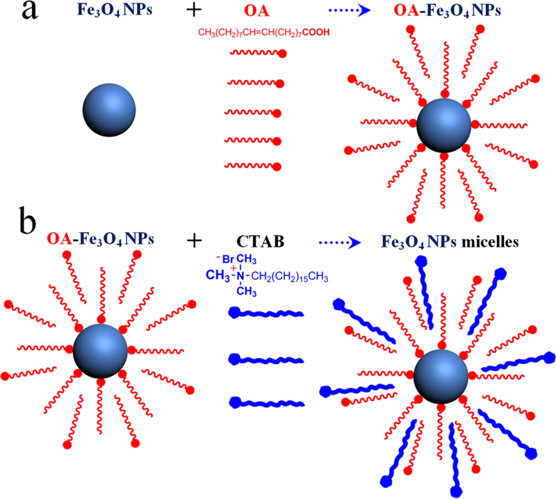
Figure 3.
Schematic illustration of Fe3O4 NP–micelles formation: (a) OA-stabilized Fe3O4 NPs; (b) Fe3O4 NP–micelles. These adopt an interdigitated bilayer structure with OA ligands as the inner layer and CTAB as the outer layer (when the OA volume is more than 30 μL, many more OA molecules adsorb onto the surface of the Fe3O4 NPs).

