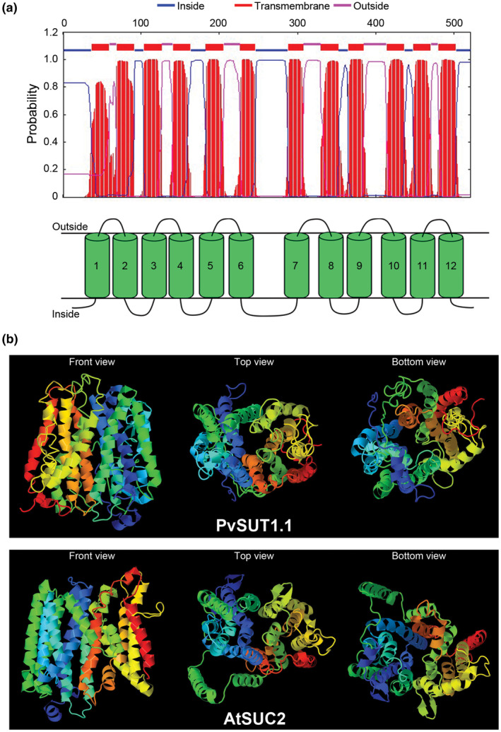
FIGURE 2.

In silico prediction analysis of PvSUT1.1. (A) Transmembrane domain prediction of PvSUT1.1 showing twelve hydrophobic regions. (B) Predicted structure of putative common bean sucrose transporter (PvSUT1.1; top panel) in comparison with the phloem localized Arabidopsis sucrose transporter (AtSUC2; bottom panel). The front, top, and bottom views of the predicted protein structure is shown in each panel
