. 2020 May 11;44(4):509–528. doi: 10.4093/dmj.2020.0058

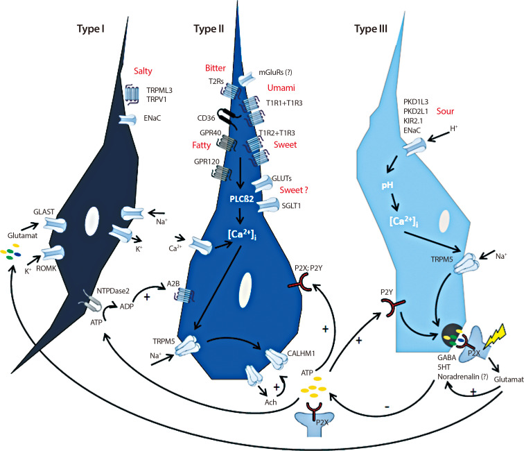
Fig. 2. Schematic presentation of cell signaling within taste bud cells. Type II cells express G-protein coupled receptors (GPRs) for bitter (taste receptors type 2 [T2Rs]), sweet (taste receptor type 1 member 1 [T1R1], T1R2, T1R3) and umami (T1R1+T1R3), but also GPR40 and 120 and glycoprotein 4 (also named cluster of differentiation 36, CD36) transducing the taste quality “fatty” [15]. In addition, metabotropic glutamate receptors (mGluRs) and glucose- and sodium/glucose transporters (GLUTs, SGLT1) are thought to transduce umami and sweet, respectively [27,31]. Binding of tastants to their cognate receptors increase intracellular calcium level ([Ca2+]i) which activates transient receptor potential cation channel subfamily M member 5 (TRPM5), a Ca2+/Na+ cotransporter [34]. This leads finally to an activation of the calcium homeostasis modulator protein 1 (CALHM1) which is meant to release adenosine triphosphate (ATP) [35,36]. ATP signals to afferent nerve fibers via binding to P2X receptors but also feeds back in an autocrine fashion via binding to P2X and P2Y receptors on type II cells [36]. In addition, type II cells secrete Acetylcholine (Ach) which further stimulates ATP secretion [38]. Moreover, ATP activates type III cells by binding to P2Y receptors [37]. This in turn initiates the release of neurotransmitters gamma aminobutyric acid (GABA), serotonin (5-HT) and noradrenaline into the presynaptic space as consequence of raised [Ca2+]i [25,40]. In addition, this release is mediated as a result of changes in pH through the uptake of H+ by ion channels such as polycystic kidney disease proteins 1 like 3 and 2 like 1 (PKD1L3, PKD2L1), inward rectifying K+ channel (KIR2.1) and the epithelial Na+ channel (ENaC) [41,42,43,44,45]. GABA and 5-HT activate afferent nerve fibers but feedback to type II cells in order to decrease further ATP secretion [15]. Glutamate is released by activated nerve fibers and tune the release of GABA and 5-HT, finally shutting down ATP secretion from type II cells [15]. Type I cells seem to have glia like function as they express several ion channels (inward rectifying K+ channel [ROMK], glutamate-aspartate transporter [GLAST], ENaC) which are supposed to clear ion currents [22,23,35]. Moreover, as ENaC has been identified in type I cells and this is thought to be the main receptor for the detection of low NaCl-salts, these cells may transduce salty [46]. Further channels involved in transducing salty are mucolipin 3 (TRPML3) and transient receptor potential cation channel subfamily V member 1 (TRPMV) [47]. However, their cellular localization has yet to be elucidated. In addition, the enzyme nucleoside triphosphate diphosphohydrolase-2 (NTPDase2) located on the surface of type I cells, is responsible for the degradation of ATP to adenosine diphosphate (ADP) [39]. In turn, sweet receptor expressing type II cells bind ADP by adenosine 2B receptors (A2B) which further increases sweet sensation [39]. TRPV1, transient receptor potential cation channel subfamily V member 1; PLCβ2, Phospholipase C beta 2.

Fig. 2