
Fig. 13.
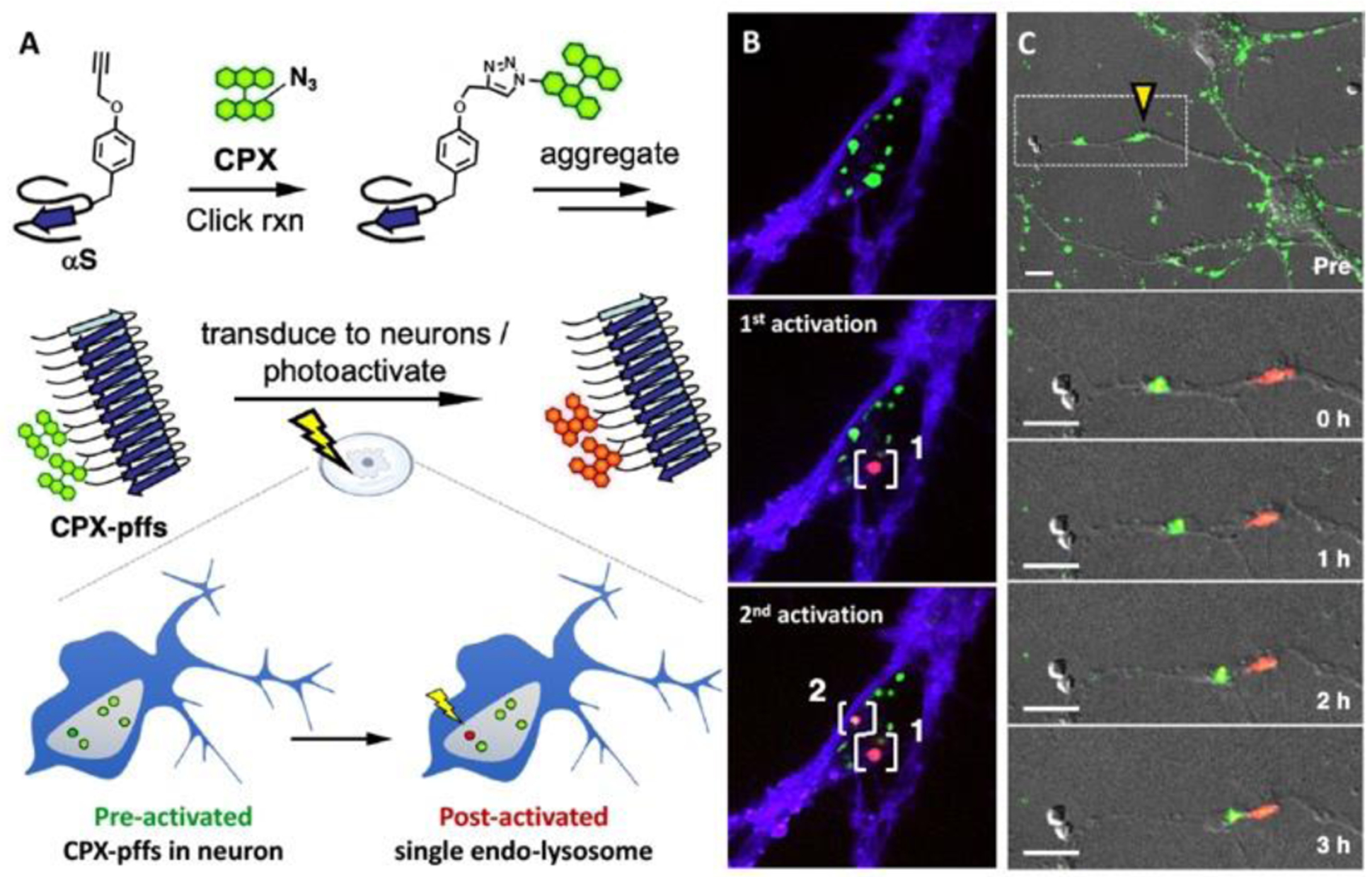
Application of photoconvertible probe (PC1) in tracking α-synuclein (αS). A. Synthesis and transduction of CPX labelled preformed fibrils of engineered αS (CPX-pffs) B. Sequential photoconversion of internalized CPX-pffs in white brackets. (blue channel: trypan blue staining extracellular membrane, green channel: unactivated CPX-pffs, and red channel: post-activated CPX-pffs) are shown in merged images. C. Tracking of CPX-pffs under yellow triangle after irradiation, zoomed in (white dotted box) below. Merged images of differential interference contrast (DIC), green and red channels are shown. Scale bars are 10 μm. Adapted from ref 85. Copyright (2019) American Chemical Society.