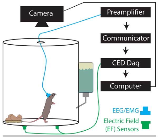
Figure 1. Cage Set-up for Electroencephalogram/Electromyogram (EEG/EMG) and Synchronized Electric Field (EF) Recordings.

The animals were singly housed in cylindrical acrylic chambers (150 oz. 8 inch diameter, 8 inches tall). Head stages (blue) were surgically mounted prior to recordings and contain EEG and EMG electrodes connected to a preamplifier, communicator, then a Cambridge data acquisition box (CED Power 1401). EF sensors (green) were attached to the exterior of the cage bottom approximately 2-3 inches from the cage wall and connected directly to the Power 1401.
