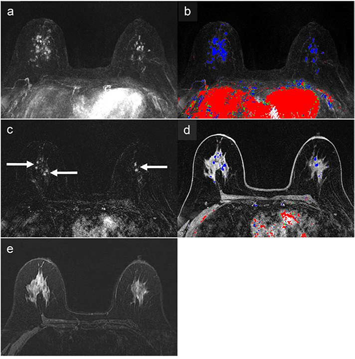
Figure 2:
67-year-old woman with a 41% lifetime breast cancer risk based on family history, as determined by the Gail model, and a variant of unknown significance in the PALB2 gene. The abbreviated breast MRI (AB-MRI) first post-contrast subtraction MIP (AB1) (a) with time-intensity color map (b) shows multiple areas of enhancement (right greater than left) in both breasts with associated persistent delayed kinetics indicated by predominantly blue coloration on the color map. The AB-MRI first post-contrast subtraction image (AB2) (c) shows a representative area centrally with multiple bilateral nonmass enhancements (arrows), right greater than left. The corresponding AB-MRI first post-contrast T1-weighted image (AB2) with a color map overlay shows associated delayed persistent kinetics (d). There was no associated increase in T2 signal on the AB-MRI T2-weighted image (AB3) (e). Both SOC-BMRI and AB-MRI exams were classified as BI-RADS 4, suspicious. MRI-guided biopsy of the right breast showed low-grade cribriform ductal carcinoma in situ (DCIS) without comedonecrosis and lobular carcinoma in situ with pagetoid extension. Subsequent right mastectomy showed cribriform atypical ductal proliferation similar to the DCIS in the MRI-guided biopsy, as well as atypical lobular hyperplasia and radial scar. Left breast MRI-guided biopsy showed a single minute focus of atypical ductal hyperplasia, as well as usual ductal hyperplasia, columnar cell change, and sclerosing adenosis. The left breast is undergoing imaging follow-up, showing benign results.

