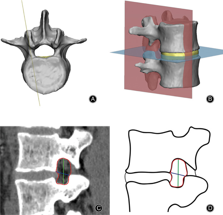
Figure 2.

Pedicle–pedicle method for measurement of the lumbar intervertebral foramen. (A) The special sagittal slice was aligned along the midline of superior and inferior pedicles. (B) The slice was perpendicular to the disc space. (C) Foraminal area (red) was defined as the area bounded by the adjacent superior and inferior vertebral pedicles, the posterosuperior portion of the inferior vertebral body, the surface of the intervertebral disk posteriorly, the posteroinferior portion of the superior vertebral body, and the surface of the ligamentum flavum anteriorly. Foraminal height (green) was defined as the longest distance between the border of the superior and the inferior pedicle. Foraminal width (blue) was defined as the distance between the posteroinferior edge of the superior vertebrae and the anterior boundary of superior articular process. (D) Diagram of foraminal area (red), foraminal height (green), and foraminal width (blue).
