
Figure 2.
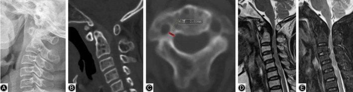
Preoperative images of the illustrative case. (A) Lateral radiograph showed slight deformity of the odontoid process. (B) CT on sagittal plane showed that the thickening of ligaments resulted in spinal stenosis. (C) CT on transverse plane showed excessive narrowness of the pedicles of C2. (D, E) MRI demonstrated severe spine canal stenosis and compression of the spinal cord.