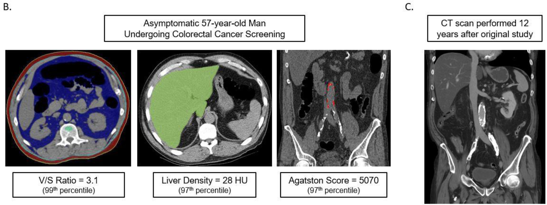
Figures 1B and 1C. Case example in an asymptomatic 57-year-old man undergoing CT for colorectal cancer screening.

At the time of CT screening, he had a BMI of 27.3 and FRS of 5% (low risk). However, several CT-based metabolic markers were indicative of underlying disease (B), including a visceral-to-subcutaneous fat ratio of 3.1 (99th percentile), abdominal aortic Agatston score of 5070 (97th percentile), and steatotic liver density of 28 HU (97th percentile). Multivariate Cox model prediction based on these three CT-based results put risk of CV event within 2, 5, and 10 years at 19%, 40%, and 67%, respectively, and of death at 4%, 11%, and 27%, respectively. At longitudinal clinical follow-up, the patient suffered an acute MI three years after this initial CT and died 12 years after CT at the age of 64. Contrast-enhanced CT (C) performed seven months before death for minor trauma was interpreted as negative but does show significant progression of vascular calcification, visceral fat, and hepatic steatosis.
