Figure 5.

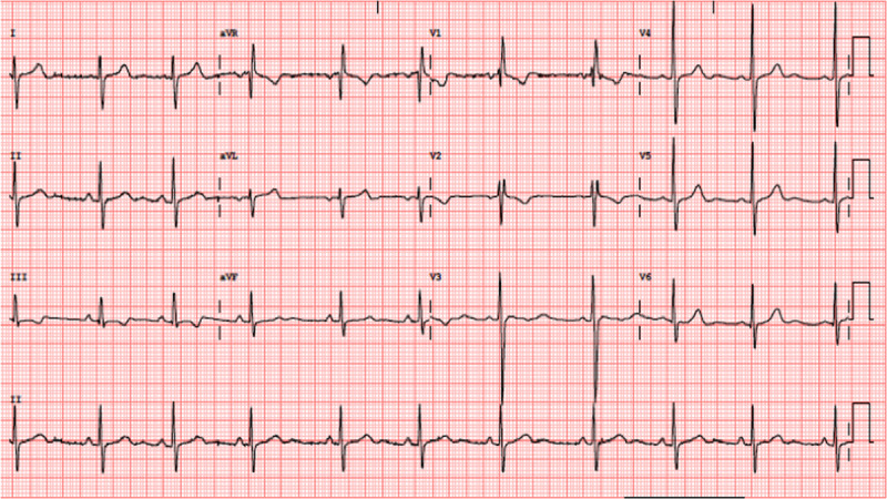
ECG performed before discharge. There is a normalization of the cQT interval and subtle changes of T wave in precordial and inferior leads.

ECG performed before discharge. There is a normalization of the cQT interval and subtle changes of T wave in precordial and inferior leads.