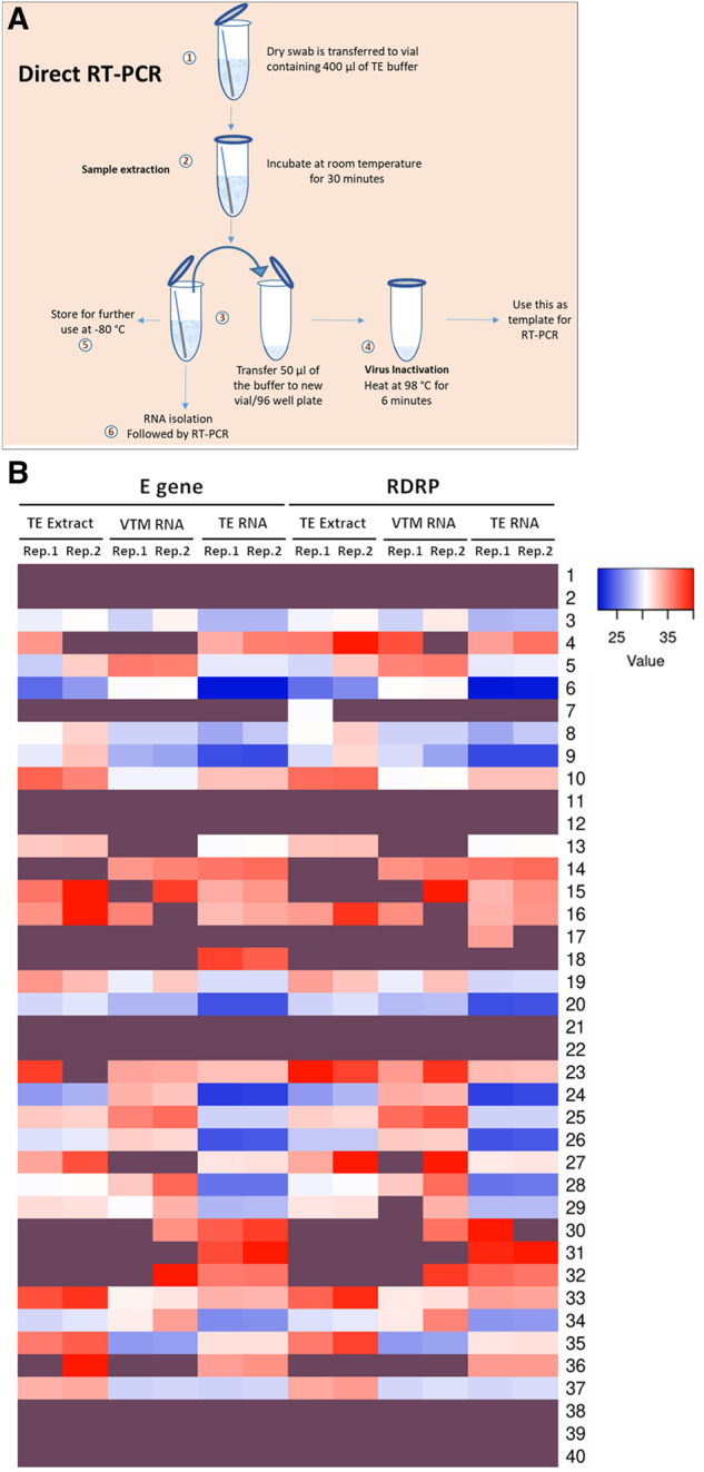
Figure 1:

RNA extracted from TE buffer outperform other methods. (A) Schematic of the entire protocol for TE-based sample extraction and RT-PCR. (B) Heatmap representing the CT values of E and RdRP genes obtained in two replicates (Reps.1 and 2) of RT-PCR using TE extract, VTM-extracted RNA (VTM-RNA), and TE-extracted RNA (TE-RNA) as templates (n = 40). Details in Supplementary data, Table S2. Samples D1–D40 are represented as 1–40. Dark purple shade represents no signal detection.
