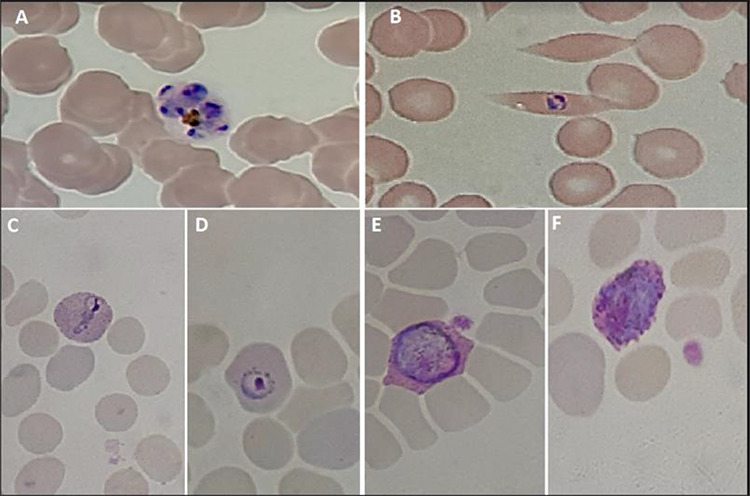
Figure 1.

Giemsa-stained thin blood smears from Case 2 (A, B) and Case 1 (C, D, E, F). (A) Typical P. ovale schizont with 4–12 merozoites and a pigment concentrated mass. (B–D) P. ovale trophozoites parasitizing enlarged (C, D) and often distorted (B) erythrocytes. (E, F) P. ovale male (E) and female (F) round gametocytes. Note: Schüffner’s dots, typical of P. ovale and P. vivax, are clearly visible in parasitized erythrocytes of Case 1
