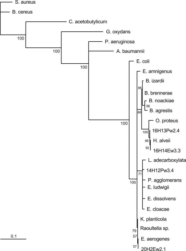
Fig 9. Phylogenetic analysis of 16S rRNA sequences (cluster II; enteric bacteria).
Complementary DNA was amplified from aerobic bacteria isolated from the gut of nurse bees (Apis millifera) from Australia, sequenced, and sequences were aligned using the Fast Fourier Transform (MAFFT). A maximum likelihood phylogenetic tree was generated using PhyML3.0 and the general time reversible (GTR) evolutionary model. Bootstrap values detected for 100 replicates are shown near the nodes. The scale bar represents the change in nucleotides of the sequence (i.e., genetic variation for the length of scale).

