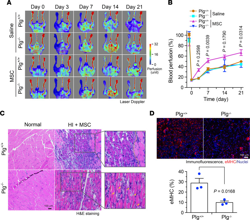
Figure 2. Plg is critical for MSC-mediated tissue repair after HI.
HI was induced in Plg+/+ and Plg−/− mice by ligation of the femoral artery, followed by MSC transplantation or saline injection in the ischemic limbs. (A and B) Mice were subjected to analysis by laser Doppler scanning. (A) Representative images are shown. (B) Blood perfusion rates in ischemic limbs were quantified and expressed as the percentage of normal limbs (n = 6–8, mean ± SEM). Statistical analysis by 2-way ANOVA (comparing MSC-treated Plg+/+ and Plg−/− mice). (C) Gastrocnemius muscle in normal or ischemic limbs was collected day 7 after the surgery and subjected to H&E staining. Arrows or left area of dashed line indicate newly regenerated muscles. Original magnification, ×100. (D) Gastrocnemius muscle in ischemic limbs was collected at day 7 after HI surgery. Tissue sections were probed with eMHC antibody. Upper, representative images. Bottom, quantitative analysis (n = 3, mean ± SEM). Statistical analysis by unpaired Student’s t test.

