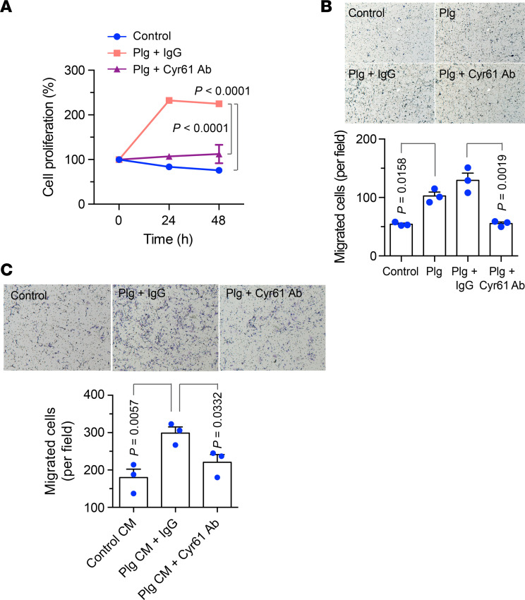
Figure 6. Plg activates Cyr61 by cleavage to stimulate cell proliferation and migration.
(A) MSCs were cultured with serum-free medium and treated with or without 20 μg/mL Plg plus IgG or Plg plus anti-Cyr61 antibody under hypoxia (2% O2). Cell proliferation was determined by MTS-based assay (n = 3, mean ± SEM). Statistical analysis by 1-way ANOVA. (B) MSCs were treated with PBS, Plg, Plg plus IgG, or Plg plus anti-Cyr61 antibody. Plg-depleted FBS was used as a chemoattractant. Upper, representative images. Bottom, quantitative analysis (n = 3, mean ± SEM). Original magnification, ×100. Statistical analysis by 2-way ANOVA. (C) ECs were treated with control CM, Plg CM plus IgG, or Plg CM plus anti-Cyr61 antibody. Plg-depleted FBS was used as the attractant to induce the migration through matrix. Upper, representative images. Bottom, quantitative analysis (n = 3, mean ± SEM). Original magnification, ×100. Statistical analysis by 1-way ANOVA. Experiments were repeated 3 times and representative results are shown.

