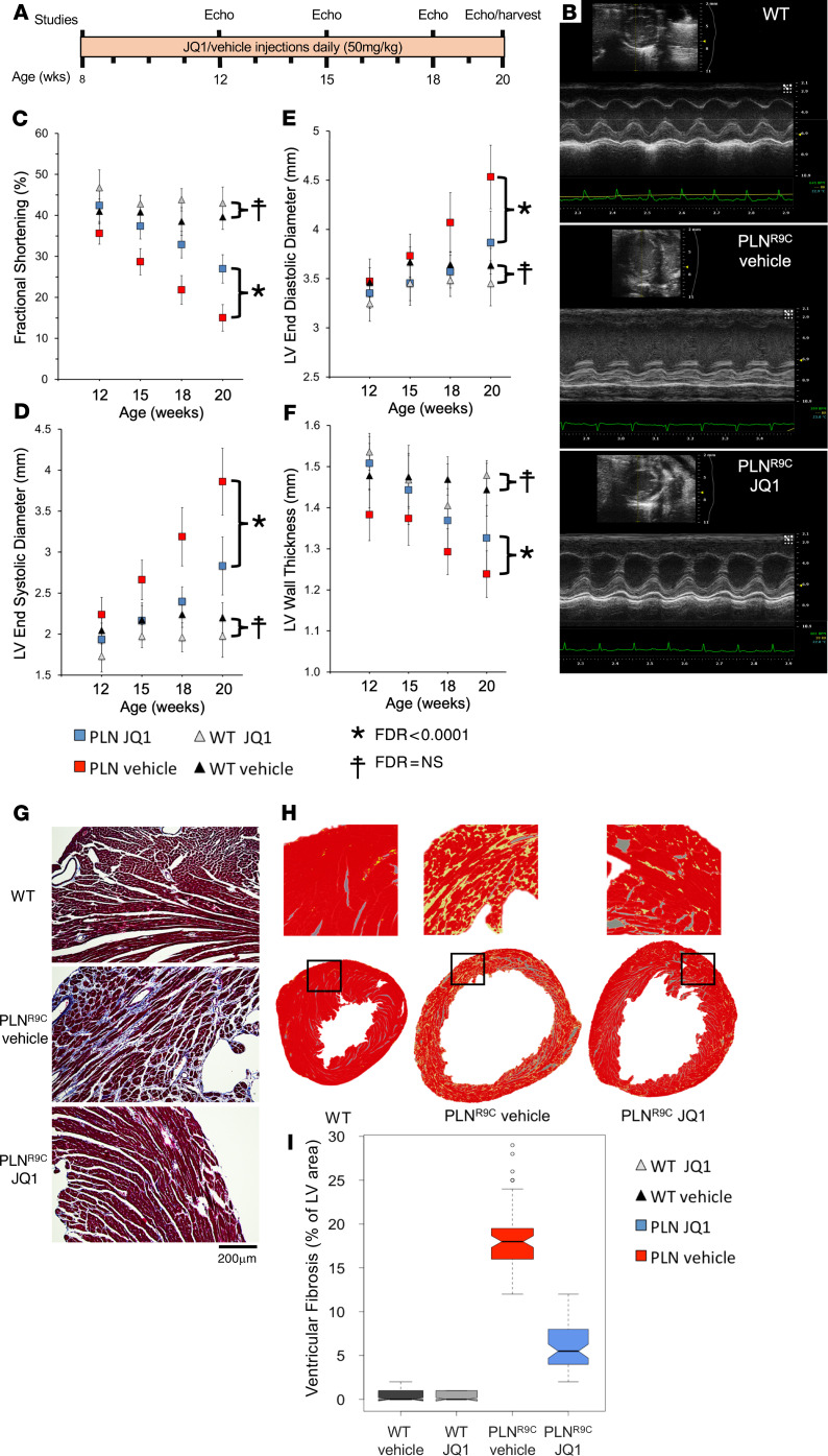
Figure 2. BET inhibition delays DCM in PLNR9C mice.
(A) Experimental protocol. (B) Representative M-mode images of WT and PLNR9C vehicle- or JQ1-treated mice at 18 weeks of age. Echo, echocardiography. (C–F) Echocardiographic assessment of mice treated with JQ1 or vehicle demonstrates progressive systolic dysfunction and negative LV remodeling in PLNR9C mice that was significantly blunted by JQ1 (n = 14 PLNR9C, n = 7 WT mice per group; ANOVA corrected for multiple hypothesis testing). Representative images demonstrating cardiac fibrosis: (G) Masson’s trichrome–stained LV sections from WT and PLNR9C hearts and (H) false-color images of fibrosis identified by Keyence microscope software (yellow, fibrosis; box, enlarged region). As JQ1 had no effect on fibrosis in WT, a single representative WT image is shown. Scale bar in G: 200 μm. Original magnification in H, ×10.(I) Quantification of scar area demonstrated severe fibrosis in PLNR9C vehicle-treated hearts at 20-weeks of age that was markedly blunted by JQ1 (n = 3 mice, 36 images from n = 4 levels from apex to base for each mouse). Boxes, IQR; whiskers, 1.5× IQR; black line, median; notches, SD, circles, extreme outlier values).

