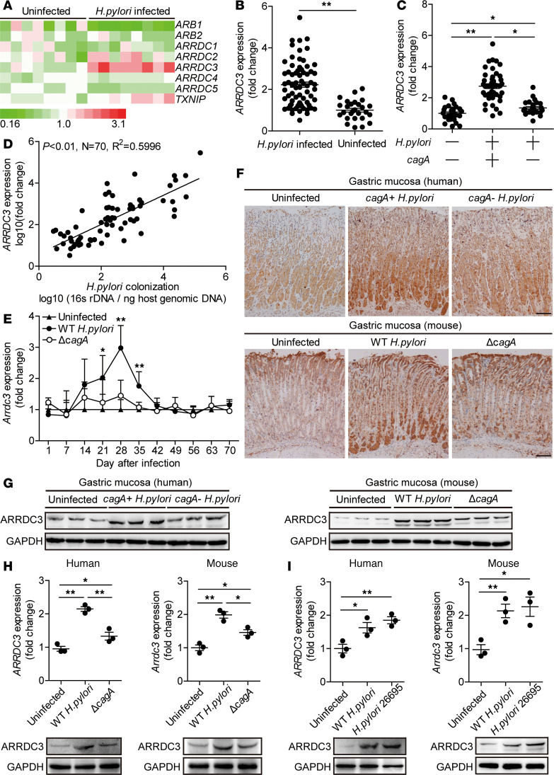
Figure 1. ARRDC3 is increased in gastric mucosa of H. pylori–infected patients and mice.
(A) The mRNA expression profiles of arrestin family members in human primary gastric mucosa of H. pylori–infected patients (n = 8) and uninfected donors (n = 8) was analyzed by real-time PCR. (B) ARRDC3 expression in gastric mucosa of H. pylori–infected (n = 70) and uninfected donors (n = 27) was compared. (C) ARRDC3 expression in gastric mucosa of cagA+ H. pylori–infected (n = 44), cagA– H. pylori–infected (n = 26), and uninfected donors (n = 27) was compared. (D) The correlation between ARRDC3 expression and H. pylori colonization in gastric mucosa of H. pylori–infected patients was analyzed. (E) Dynamic changes of Arrdc3 expression in gastric mucosa of WT H. pylori–infected, ΔcagA-infected, and uninfected mice. n = 5 per group per time. (F and G) ARRDC3 protein in gastric mucosa of cagA+ H. pylori–infected, cagA– H. pylori–infected, and uninfected donors or in gastric mucosa of WT H. pylori–infected, ΔcagA-infected, and uninfected mice on day 28 p.i. was analyzed by immunohistochemical staining (F) and Western blot (G). Scale bars: 100 μm. (H) ARRDC3/Arrdc3 expression in human primary gastric mucosa from uninfected donors/mice infected with WT H. pylori or ΔcagA ex vivo analyzed by real-time PCR and Western blot (n = 3). (I) ARRDC3/Arrdc3 expression in human primary gastric mucosa from uninfected donors/mice infected with WT H. pylori or H. pylori 26695 ex vivo analyzed by real-time PCR and Western blot (n = 3). Data are representative of 2 independent experiments. Data are mean ± SEM and analyzed by Student t test, Mann-Whitney U test, and 1-way ANOVA. Western blot results are run in parallel and contemporaneously. *P < 0.05, **P < 0.01 for groups connected by horizontal lines or compared with uninfected mice.

