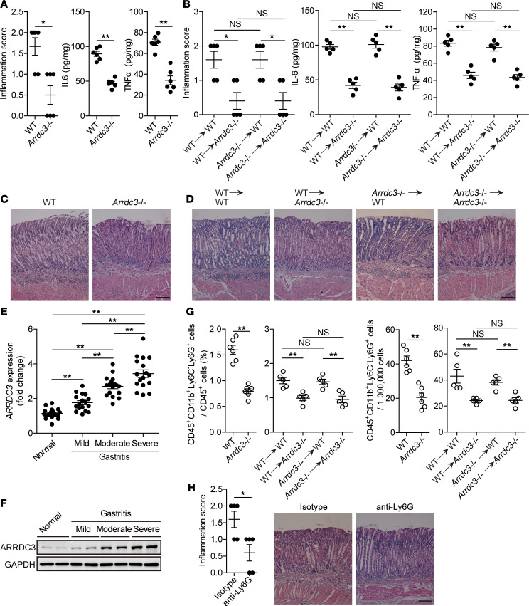
Figure 4. ARRDC3 has proinflammatory effects during H. pylori infection.
(A and B) Histological scores of inflammation and IL-6 and TNF-α protein in gastric mucosa of WT H. pylori–infected WT and Arrdc3–/– mice (n = 6) (A) or in gastric mucosa of WT H. pylori–infected BM chimera mice (n = 5) (B) on day 28 p.i. were compared. (C and D) Representative H&E staining images showed inflammation in gastric antra of WT H. pylori–infected WT and Arrdc3–/– mice (C) or in gastric antra of WT H. pylori–infected BM chimera mice (D) on day 28 p.i. Scale bars: 100 μm. (E and F) ARRDC3 expression (E) and ARRDC3 protein (F) in gastric mucosa of H. pylori–infected patients with mild (n = 17), moderate (n = 17), severe inflammation (n = 17), and uninfected donors with normal gastric histopathology (n = 19) was compared. (G) CD45+CD11b+Ly6C–Ly6G+ neutrophil levels in gastric mucosa of WT H. pylori–infected WT and Arrdc3–/– mice (n = 6) or in gastric mucosa of WT H. pylori–infected BM chimera mice (n = 5) on day 28 p.i. were compared. (H) Histological scores of inflammation in gastric mucosa of WT H. pylori–infected mice injected with Abs against Ly6G or corresponding isotype control Abs on day 28 p.i. were compared (n = 5). Scale bars: 100 μm. Data are representative of 2 independent experiments. Data are mean ± SEM and analyzed by Student t test, Mann-Whitney U test, and 1-way ANOVA. Western blot results are run in parallel and contemporaneously. *P < 0.05, **P < 0.01 for groups connected by horizontal lines.

