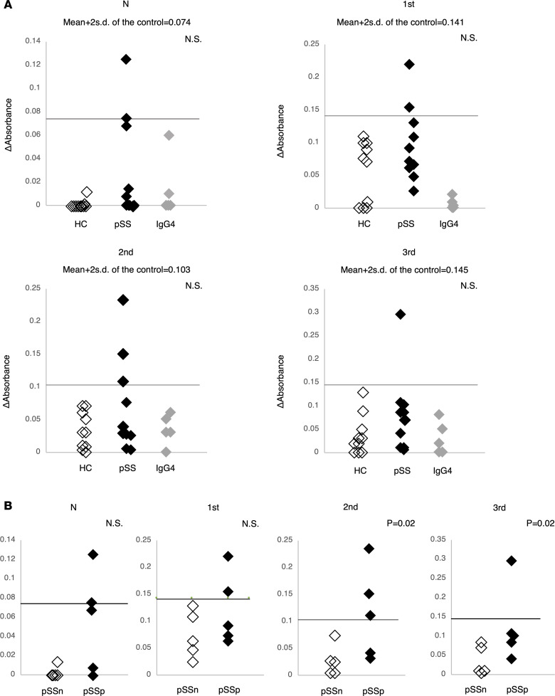
Figure 6. Anti-M3R antibodies against 4 extracellular domains of M3R.
Anti-M3R antibodies against extracellular domains of M3R were examined as described previously by ELISA (12). The cutoff level between negative and positive values of each anti-M3R antibody represented the mean of the normal controls + 2 SD values, represented by the horizontal line in each figure. (A) The frequency and titers of anti-M3R antibody were not significantly different among HS, IgG4-RD, and pSS. Only pSS were positive for anti-M3R antibodies. *P < 0.05, by Kruskal-Wallis test. (B) Antibody titers against second and third extracellular domains were significantly higher in the 5 pSS patients positive for M3R-reactive Th17 cells (P = 0.02, each). *P < 0.05, by the Mann-Whitney U test. N, N terminal; 1st, first extracellular loop; 2nd, second extracellular loop; 3rd, third extracellular loop; pSSn, M3R-reactive Th17–negative patients; pSSp, M3R-reactive Th17–positive patients.

