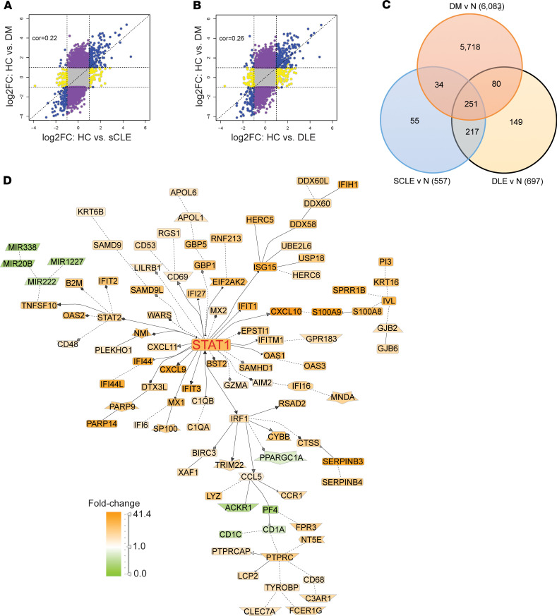
Figure 3. DM shares IFN genes with CLE lesions.
(A and B) Comparison of DEGs in DM lesional skin (y axis) versus DEGs in SCLE (A) and DLE (B). Shared DEGs in the same direction are denoted in blue. (C) Venn diagram showing shared and unique DEGs between DM, SCLE, and DLE. (D) Genomatix Pathway analysis of shared 251 DEGs between DM and CLE subtypes.

