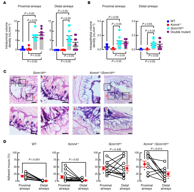
Figure 5. Genetic silencing of Kcnn4 improved mucus clearance in mouse airways.
Intraluminal (A) and intracellular (B) mucus volume was determined in the proximal and distal airways. Differences were calculated with ANOVA on ranks; n = 12, 12, 12 and 9 animals for WT, Kcnn4+/+, Scnn1btg/+, and double mutants, respectively. Representative images of mucus attachment to the epithelial surface for Scnn1btg/+ (n = 15) and double mutants (n = 13) (C). Selected areas of main images (scale bar: 50 μm) are noted as 1–3 in red letters, and are shown amplified separately (scale bar: 20 μm). Summary of the percentage of epithelium surface covered my mucus in proximal and distal airways (D). Only paired samples from the same animal were included; n = 13, 16, 15, and 13 for WT, Kcnn4+/+, Scnn1btg/+, and double mutants, respectively. *P < 0.05 vs. WT and Kcnn4–/– ANOVA on ranks. The P values for each proximal vs. distal airways comparison were calculated by rank-sum test.

