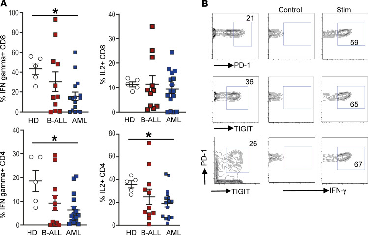
Figure 2. Changes in BM T cell function in children with B-ALL and AML at diagnosis.
BMMNCs from B-ALL (n = 13), AML (n = 17), or HD (n = 5) were cultured alone or with PMA/ionomycin in the presence of GolgiStop. After 4 hours of culture, cells were stained with dead cell exclusion dye as well as antibodies to detect surface CD3, CD4, CD8, PD-1, TIGIT, intracellular IFN-γ, and IL-2 and analyzed using flow cytometry. (A) Proportion of CD8+ and CD4+ T cells secreting IFN-γ and IL-2. (B) IFN-γ secretion by cells expressing PD-1 and/or TIGIT. Figure shows a representative plot from patient with AML. All graphs show mean ± SEM. *P < 0.05 by Mann-Whitney U test with Bonferroni’s correction for multiple comparisons.

