
Fig. 6. PS3D1@DMXAA inhibits breast tumor metastasis and synergizes with ICB to inhibit B16-melanoma.
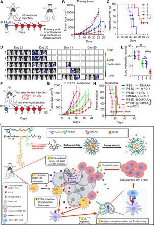
(A) Schematic diagram of the orthotopic breast tumor model and administration method. Mice bearing 4T1-Luci breast tumors were treated as in Fig. 5A. (B) Tumor growths are shown (n = 8). (C) Survival curves were compared using log-rank test (n = 8). (D) In vivo bioluminescence images of 4T1-Luci lung metastatic tumors. (E) Number of the lung metastatic nodules on day 25. One-way ANOVA with Tukey’s test. (F) Combined treatment scheme for mice with established B16.F10 tumors. (G) Tumor growths of B16.F10 tumor-bearing mice are shown (n = 8, means ± SEM). (H) Survival curves were compared using log-rank test (n = 8). Data are means ± SD, and statistical significance was calculated by two-tailed Student’s t test unless otherwise indicated. ***P < 0.001 and **P < 0.01. (I) Schematic illustration of the self-assembly of PS3D1@DMXAA nanoparticles with redox-responsive drug release in tumor cells. The electrostatic interaction between the tertiary amine group and DMXAA provides efficient drug loading. (1 and 2) Redox stimuli trigger SN38 release in tumor cells. SN38 induces tumor cell death and release of chemokine CCL4 that drives the infiltration of CD103+ DCs in the TME. (3) Meanwhile, PS3D1@DMXAA elicits efficient cytosolic delivery of DMXAA for STING activation in CD103+ DCs. Together, these enhance the maturation and TAA uptake of CD103+ DCs. (4 and 5) STING activation enhances the migration of mature CD103+ DCs into the tdLN and stimulates TAA cross-presentation by CD103+ DCs for cross-priming of TAA-specific effector CD8+ cytotoxic T cells. (6) PS3D1@DMXAA modulates the immunosuppressive TME and facilitates TAA-specific effector CD8+ cytotoxic T cell recruitment through CXCL9/CXCL10. All these eventually amplify the antitumor therapeutic effects.