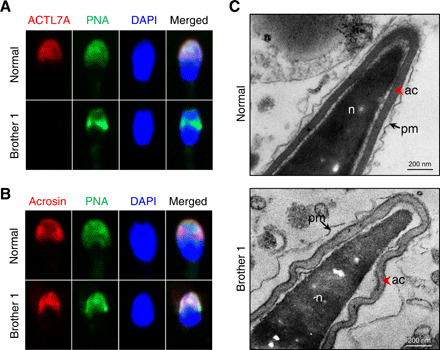
Fig. 2. Homozygous mutation in ACTL7A caused the ultrastructural impairment of sperm acrosomes in the two infertile brothers.

(A) The signal of ACTL7A disappeared in the case of the spermatozoa from the infertile brothers carrying the homozygous ACTL7A mutation. The localization of ACTL7A was identified using polyclonal antibodies against ACTL7A, followed by the secondary antibodies (donkey anti-rabbit–Cy3; red). The acrosome and sperm nuclei were stained with PNA (green) and DAPI (blue), respectively. Scale bars, 2 μm. (B) The acrosome signals were distributed nonuniformly in sperm of brother 1. Acrosin (red) was a marker of acrosomal matrix. PNA (green) labels the outer acrosomal membrane. (C) The ultrastructure of sperms from the brothers with the ACTL7A mutation revealed acrosome detachment. The red arrowhead indicates the acrosome. The arrow indicates the plasma membrane. ac, acrosome; n, nucleus; pm, plasma membrane. Scale bars, 200 nm. Photo credit: Aijie Xin, Fudan University.
