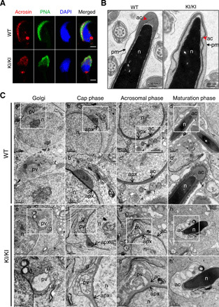
Fig. 4. Homozygous knock-in mutation in mouse Actl7a causes acrosomal defects due to fusion failure of proacrosomal vesicle.

(A) The location of acrosome changed in sperm from the Actl7aKI/KI mice. Acrosin (red) and PNA represented acrosomal matrix and the outer acrosomal membrane, respectively. Sperm nuclei were stained with DAPI (blue). Scale bars, 2 μm. (B) TEM analysis revealed the detachment of the acrosome (ac) from the sperm nuclei (n) in the sperms from the Actl7aKI/KI mice. The red arrowhead indicates the acrosome. The arrow indicates the plasma membrane (pm). (C) Acrosome biogenesis of WT and Actl7aKI/KI mouse spermatid. (a to d) Four phases of acrosome biogenesis in WT. Golgi phase (a), cap phase (b), acrosomal phase (c), and maturation phase (d). (a′ to d′) Higher magnification of the boxed region from (a) to (b). Numerous proacrosomal vesicles derived from the trans-face of the Golgi (a and a′) attached to the acroplaxome forming a cap over the nucleus (b and b′). Acrosome further developed and flattened over half of the nucleus in acrosomal phase (c and c′) and continued to maturation (d and d′). (e to e′) Four corresponding phases in Actl7aKI/KI mouse. (e′ to h′) Higher magnification of the boxed region from (e) to (h). Big and atypical proacrosomic vesicles generated during Golgi phase (e and e′). The proacrosomal vesicle fusion failed (f and f′) and accumulated gradually (g and g′), eventually causing the abnormal acrosome detached from the nuclear envelope (h and h′). pv, proacrosomal vesicles; g, Golgi; n, nucleus; ag, acrosomal granule; apx, acroplaxome. Photo credit: Aijie Xin, Fudan University.
