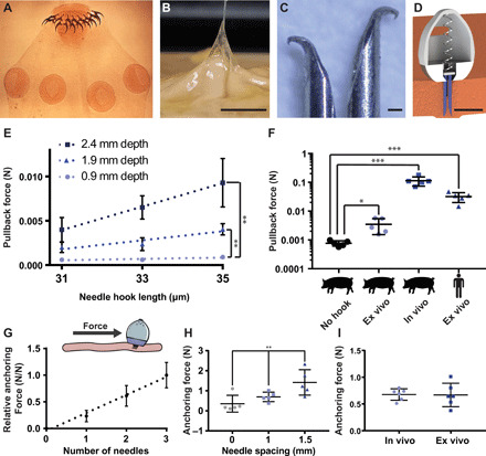
Fig. 2. Implanted hooked needles generate retention forces that allow devices to remain localized to the stomach wall for prolonged time periods.

(A) Scolex of the Taenia solium tapeworm, a parasitic worm that uses hooks to attach to the GI tract of its host. For scale, the diameter of a typical scolex for this species is 1 mm. Photo credit: Centers of Disease Control, public domain. (B) Hooks at the end of a 32-gauge needle as small as 30 μm in size latched onto ex vivo human stomach tissue. Scale bar, 5 mm. Photo credit: Alex Abramson, MIT. (C) Hooked needles fabricated in different sizes and combined into arrays created an attachment mechanism similar to the tapeworm, Scale bar, 100 μm. Photo credit: Alex Abramson, MIT. (D) The STIMS device inserted arrays of hooked needles into the stomach tissue. A 3D CAD model of the STIMS device, Scale bar, 5 mm. (E) Pullback forces from hooked needles inserted into ex vivo swine stomach (n = 3). (F) Pullback forces from hooked needles inserted into swine and human stomach tissues (n = 5). “No hook” data represent frictional pullback force from ex vivo swine tissue when using needles that do not hook onto the tissue. (G) The relative anchoring force was linearly correlated with the number of needles inserted into tissue (n = 9 over three stomachs). (H) Increasing the distance between the inserted needles increased the anchoring force (n = 5). (I) Anchoring forces were on average 0.7 N in both in vivo and ex vivo swine stomachs when using a STIMS device with three needles spaced 1 mm apart (n = 6 over two stomachs). Means ± SD; *P < 0.05; **P < 0.01; ***P < 0.001.
