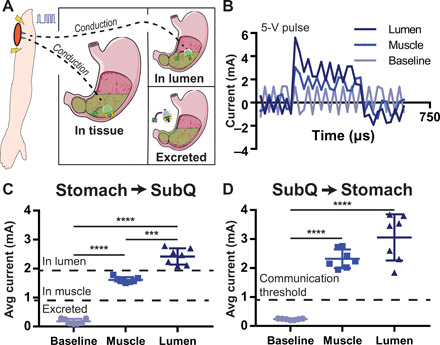
Fig. 5. In vivo conductive communication conveys tissue wall localization.

(A) The conductive nature of the body allows electrical pulses to pass through the tissue from the stomach to the subcutaneous space. (B) Current readout from a circuit branch containing a 10-ohm resistor connected to the subcutaneous space of a swine when a 330-μs, 5-V electrical pulse is generated by a microcontroller in the stomach. Electrical pulses have different profiles depending on if they originate in the stomach lumen or stomach muscle. (C) Average current of the pulse passing through the body in the subcutaneous space when a pulse is generated in the stomach. Depending on the current reading, it is possible to discern whether probes are in the stomach muscle, are floating in GI luminal fluid, or are not in contact with the body. (D) Pulses generated in the subcutaneous space can be read in the stomach as well. Means ± SD; ***P < 0.001; ****P < 0.0001.
