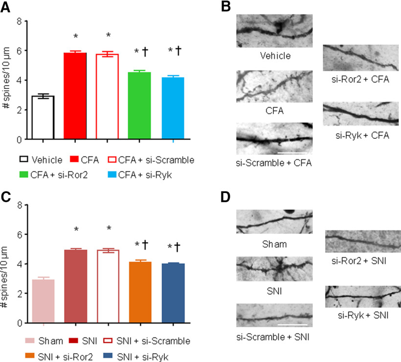
Figure 8.
Ror2 and Ryk contribute to spine plasticity in spinal neurons in chronic pain models. A, C, Quantification of synaptic spine density in spinal dorsal horn neurons in vivo 24 h after the injection of CFA (A) or 7 d after SNI nerve lesion (C) in the presence or absence of intrathecal administration of a siRNA against Ror2 or Ryk. B, D, Examples of high-magnification views of microscopic images of labeled dendrites with synaptic spines are shown for the groups represented in A and C. N = 3–4 animals/condition, 20–30 neurons, 500–700 spines counted and analyzed. Two-way ANOVA for random measurements, followed by Bonferroni's test, were performed. *p < 0.05 compared with control group (vehicle or sham treated); †p < 0.05 compared with the group that received only the nociceptive stimuli but not the inhibitor. In all panels, data are represented as the mean ± SEM. Scale bars: B, D, 10 μm.

