Figure 1.
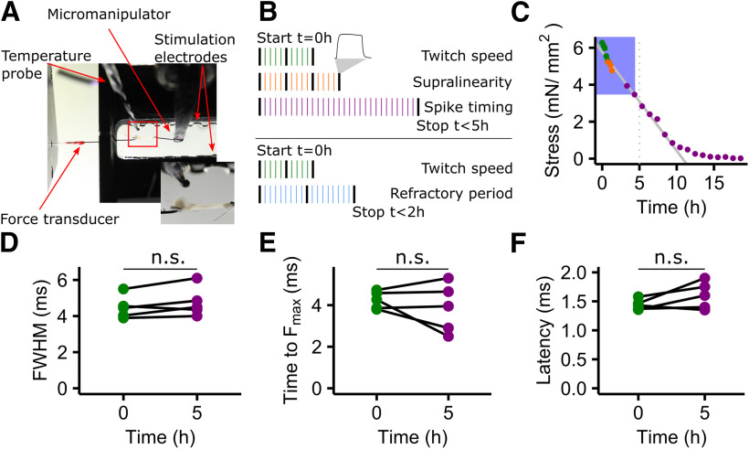
Muscle speed does not change over experimental time. A, In vitro muscle physiology setup. Muscle fiber bundles were fixed to a force transducer (left) on the rostral end and to a motorized micromanipulator (right) on the caudal end. The bath chamber (black) was continuously supplied with fresh oxygenated Ringer's solution. The red box indicates the inset showing a closeup of a muscle fiber bundle in the setup. B, Schematic of the experimental timeline and design. Two sets of experiments (1 − 3 and 1 + 4) were run on two different groups of animals. One iteration consisted of a 50 ms high-frequency control stimulation (black bars) and several different stimulation patterns (colored bars). Stimulation patterns were assigned randomly within each iteration. C, Maximal tetanic force declines over time. Depicted is an example of one muscle preparation. The decline is linear (solid gray line) over >5 h (dotted vertical line). D–F, Data included in this study were all acquired within 5 h (blue box), and over this time twitch kinematics did not change significantly (paired t test; FWHM: t = −1.917, df = 4, p = 0.13, time to Fmax: t = 0.920, df = 4, p = 0.41; latency: t = −1.805, df = 4, p = 0.15). The coloring of all dots is according to B.

