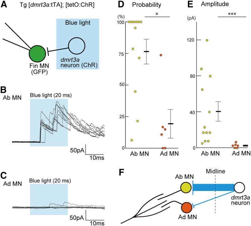
Figure 6.
dmrt3a neurons form strong inhibitory synaptic connections onto abductor MNs. A, Experimental scheme. ChR was expressed in dmrt3a neurons using the Tet system. Whole-cell voltage-clamp recording with the holding potential at +10 mV was made for an abductor or adductor MN. A 20 ms pulse of blue light illumination was applied near the region surrounding the recorded neuron (covering muscle segments 3.5–5.5). B, An example whole-cell recording from an abductor MN. Eight traces are superimposed. C, Same as B, but for adductor MNs. D, Connection probability from dmrt3a neurons to abductor or adductor MNs. Each circle represents one cell (n = 12 for abductor MNs; n = 6 for adductor MNs). *p < 0.05 (Mann–Whitney U test, p = 0.013). E, Amplitude of the peak currents recorded in abductor or adductor MNs. The amplitude presented is the mean of the peak currents in the trials for each cell. Each circle represents one cell (n = 12 for abductor MNs; n = 6 for adductor MNs). ***p < 0.005 (Mann–Whitney U test, p = 0.0017). F, A schematic illustration of the connections from dmrt3a neurons to abductor or adductor MNs.

